Neurotechnology · Computational neuroscience

Christoph Dinh

My work focuses on neural signal acquisition, real-time processing and decoding, with applications in medical technology and brain–computer interfaces.

It runs from methods research on real-time source imaging, through open-source scientific software, to the development of medical systems in industry.

Head of Physical AI & Computing, ZEISS

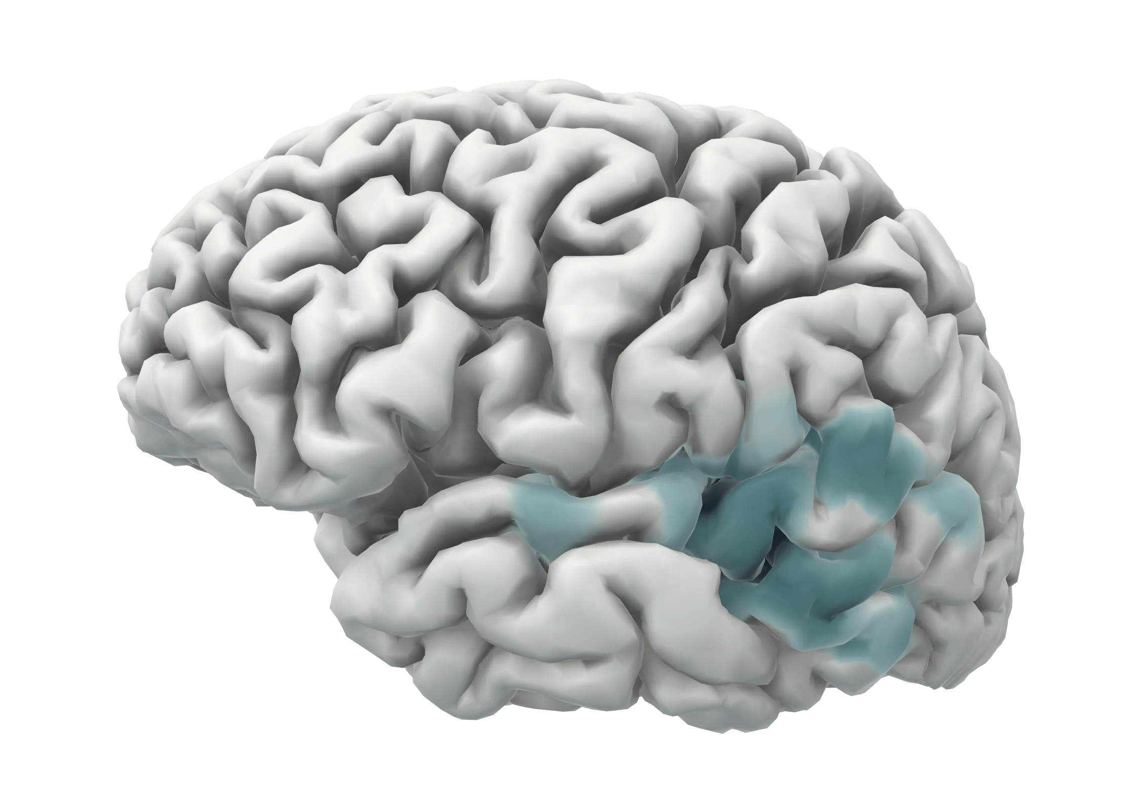
Left hemisphere of a human cortex. A shaded area around the Sylvian fissure and the upper temporal lobe marks where the source estimate is strongest.

Fig. 1Latency 92 ms

Tone100 ms200 ms

Auditory evoked response. Source estimate (dSPM) of magnetoencephalography recorded while tones were played to the right ear, shown on the cortical surface of the participant, left hemisphere. The trace is the root-mean-square signal of the planar gradiometers, peaking at 42 fT/cm.

dSPM 5–18MNE sample dataset, average of 61 trials. Computed with MNE-Python.

Research

In detail

Three connected areas. The first is established work with a published record, the third continues it, and the second is the direction I am moving towards.

  1. 01Established work

    Neural sensing and source imaging

    Magneto- and electroencephalography follow brain activity with millisecond resolution, but locating its sources is an ill-posed inverse problem that is normally solved after the recording. My doctoral and postdoctoral work made source estimates available during the measurement: a reduction of the source space based on a cortical atlas, GPU implementations of scanning methods, and the acquisition software needed to run them on clinical systems.

    Brain Topogr 2015J Neurosci Methods 2018

  2. 02Research interest

    Photonic and electronic neural interfaces

    Interfaces that stimulate as well as record have to do so selectively and without corrupting the recording. Combining electrical read-out with optical methods is one candidate, and an open engineering problem: light delivery, heating, photoelectric artefacts and stability over years all have to be controlled. I am interested in how such interfaces can be modelled and evaluated before they are built. This is planned work; I report no results here.

    Outline

  3. 03Current research

    Neural decoding and closed-loop systems

    Decoding turns measured activity into something usable: a state, an intention, a control signal. My earlier work used recurrent networks to bring temporal context into source estimates. Current work concerns decoding from non-invasive recordings and the real-time systems needed to close the loop between measurement and response.

    Front Neurosci 2021NeurIPS TS4H 2025

Selected work

All work
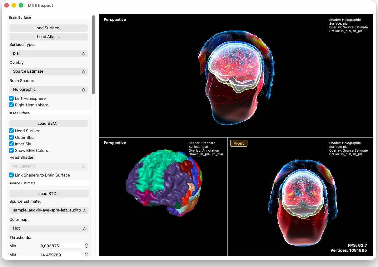
Screenshot of MNE Inspect with three 3D views of a head and brain: two with a source estimate drawn on the cortex inside a transparent head, one with the cortex coloured by atlas region.
Fig. 2MNE Inspect, the 3D viewer of MNE-CPP, showing a source estimate from the MNE sample dataset and a cortical parcellation. Screenshot from the project documentation.

Since 2010 · Open-source software

MNE-CPP

An open-source C++ framework for real-time and offline processing of MEG, EEG and related electrophysiological data: libraries for file handling, forward and inverse modelling and signal processing, and applications for acquisition, analysis and 3D visualisation. I initiated the project and co-lead its development. More than forty people have contributed to it.

  • 2017–2025

    MRI systems and ScanHub

    Acquisition software for magnetic resonance imaging: a language for pulse sequences, a receiver implemented on a graphics card, and an open platform for scanner control.

  • Since 2026

    The BCI Briefing

    An independent editorial project that follows research, clinical studies and companies in brain–computer interfaces, with a source for every claim.

  • 2015–today

    Medical-device software in industry

    Surgical navigation, neonatal MRI, cardiac mapping and platforms for machine learning in regulated products, as architect and team lead.

Selected publications

Full list

2016

Okada Y, Hämäläinen M, Pratt K, Mascarenas A, Miller P, Han M, Robles J, Cavallini A, Power B, Sieng K, Sun L, Lew S, Doshi C, Ahtam B, Dinh C, Esch L, Grant E, Nummenmaa A, Paulson D. BabyMEG: A whole-head pediatric magnetoencephalography system for human brain development research. Review of Scientific Instruments 2016;87(9):094301.

About

Portrait of Christoph Dinh

I am a biomedical engineer. I studied at Technische Universität Ilmenau and completed a doctorate there in 2015 on the real-time localisation of neuronal activity, with research stays and later a postdoctoral fellowship at the Athinoula A. Martinos Center for Biomedical Imaging at Massachusetts General Hospital and Harvard Medical School.

Since then I have developed medical systems in industry: surgical navigation at Stryker, magnetic resonance imaging at Neoscan Solutions and cardiac mapping at Ablacon. I now lead Physical AI & Computing at the ZEISS Innovation Hub @ KIT (opens in a new tab) in Karlsruhe, where I also coordinate the company’s technology field of brain–computer interfaces.